Elbow dysplasia is one of the most common causes of front leg lameness in dogs, especially in medium to large breeds. It is a complex, inherited orthopaedic disorder involving abnormal development of the elbow joint, leading to pain, stiffness, and arthritis. While the condition can be serious, early diagnosis and proper management can help affected dogs live long, active, and comfortable lives. This comprehensive guide explains everything you need to know about elbow dysplasia, from its causes and symptoms to treatment options and prevention.
What Is Elbow Dysplasia?
The elbow joint is one of the most complex joints in a dog’s body. It acts as a hinge, allowing smooth movement of the forelimb. The joint is formed by three bones:
- Humerus (upper arm bone)
- Radius (one of the forearm bones)
- Ulna (the other forearm bone)
For the joint to function properly, these three bones must fit together precisely. In dogs with elbow dysplasia, abnormal bone growth or development disrupts this alignment. The resulting uneven pressure within the joint leads to cartilage damage, inflammation, and eventually arthritis.
The Four Components of Elbow Dysplasia
Elbow dysplasia is not a single condition but rather a group of developmental abnormalities that can occur individually or together. The four main components are:
1. Fragmented Coronoid Process (FCP)
A small piece of bone on the ulna (the coronoid process) breaks off due to abnormal stress or development. This fragment irritates the joint and causes pain, inflammation, and lameness.
2. Ununited Anconeal Process (UAP)
The anconeal process, another bony projection on the ulna, fails to fuse properly with the rest of the bone during growth (typically by 20 weeks of age). This causes joint instability and chronic pain.
3. Osteochondritis Dissecans (OCD)
A defect in cartilage formation causes a flap of cartilage to separate from the underlying bone on the humerus. This leads to inflammation and restricted movement.
4. Elbow Incongruity
The joint surfaces of the radius, ulna, and humerus do not fit together evenly, leading to abnormal pressure points, cartilage wear, and progressive arthritis.
Many dogs have more than one of these abnormalities at once, making diagnosis and treatment more challenging.
Causes and Risk Factors
Elbow dysplasia is primarily a hereditary condition, but several environmental and developmental factors can worsen or trigger it.
Genetic Factors
Inherited abnormalities in bone growth are the leading cause. Certain breeds are predisposed due to genetics and selective breeding.
Breeds Commonly Affected
- Labrador Retriever
- Golden Retriever
- German Shepherd
- Rottweiler
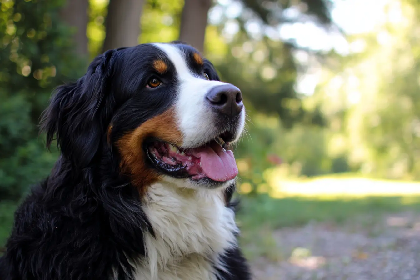
- Bernese Mountain Dog
- Newfoundland
- Saint Bernard
- Basset Hound
Environmental and Lifestyle Factors
- Rapid growth: Feeding high-calorie diets to puppies can cause bones to develop unevenly.
- Excess weight: Overweight dogs place more strain on developing joints.
- Overexercising during growth: High-impact activity before full skeletal maturity can damage joint surfaces.
- Nutritional imbalance: Diets with too much calcium or an improper calcium-to-phosphorus ratio can affect bone growth.
Symptoms and Signs
The symptoms of elbow dysplasia vary depending on the severity and which part of the joint is affected. They often begin when a dog is between 4 and 12 months old, though mild cases may not appear until adulthood when arthritis develops.
Common Symptoms Include:
- Front leg lameness: Often worse after exercise and may affect one or both legs.
- Stiffness after rest: Especially noticeable when the dog gets up in the morning.
- Reluctance to exercise or play: The dog may tire quickly or avoid running and jumping.
- Elbows held outward or rotated: Dogs may alter their stance to relieve discomfort.
- Pain when extending or flexing the elbow: The dog may yelp or resist movement.
- Swelling around the joint: Occasionally visible in more advanced cases.
- Muscle loss (atrophy): Over time, the shoulder muscles may waste away as the dog avoids using the affected leg.
If untreated, elbow dysplasia often leads to degenerative joint disease (osteoarthritis), causing chronic pain and reduced mobility.
Diagnosis
Diagnosing elbow dysplasia requires a combination of clinical evaluation and imaging studies.
Diagnostic Methods:
- Physical Examination:
- The vet assesses lameness, range of motion, and pain response in the elbow joint.
- X-rays (Radiographs):
- Standard imaging can reveal bone changes, but early cases may not show obvious abnormalities.
- CT Scan (Computed Tomography):
- Provides a detailed 3D image of the elbow, making it the gold standard for diagnosing subtle bone defects such as FCP or incongruity.
- Arthroscopy:
- A minimally invasive procedure using a small camera inserted into the joint to visualise damage directly. It can also be used for treatment.
- Genetic Testing:
- Available in some breeds to identify hereditary predisposition.
Early diagnosis is essential for effective management and to prevent irreversible joint damage.
Treatment Options
The goal of treatment is to relieve pain, restore function, and slow the progression of arthritis. The approach depends on the severity, age, and general health of the dog.
Non-Surgical (Conservative) Management
For mild cases or dogs unsuitable for surgery:
- Weight management: Keeping the dog lean reduces joint stress.
- Exercise modification: Controlled, low-impact exercise (like swimming or leash walks) helps maintain muscle tone without overloading the joints.
- Physical therapy: Hydrotherapy, massage, and specific strengthening exercises improve mobility.
- Medications:NSAIDs (non-steroidal anti-inflammatory drugs) for pain and inflammation.
- Joint supplements such as glucosamine, chondroitin, and omega-3 fatty acids to support cartilage health.
- Environmental modifications: Use ramps instead of stairs, non-slip flooring, and soft bedding.
Surgical Treatment
Surgery is often recommended for moderate to severe cases or when conservative management fails.
Common Surgical Procedures:
- Arthroscopic fragment removal (for FCP or OCD):
- The loose fragment is removed via keyhole surgery to reduce pain and inflammation.
- UAP repair or removal:
- The ununited anconeal process may be surgically reattached using screws or removed entirely.
- Sliding humeral osteotomy (SHO):
- Alters the alignment of the humerus to relieve joint pressure.
- Proximal ulnar osteotomy (PUO):
- Realigns the ulna to improve joint congruity and reduce stress.
- Elbow replacement or arthrodesis (joint fusion):
- Reserved for advanced, end-stage arthritis when other treatments are no longer effective.
Postoperative care includes rest, controlled rehabilitation, and physiotherapy to ensure optimal recovery.
Prognosis
The outlook for dogs with elbow dysplasia varies depending on the type and severity of the condition and how early it is diagnosed.
- Mild cases: With early treatment and weight management, many dogs lead normal, active lives.
- Moderate to severe cases: Surgery and lifelong management can provide good pain control and function.
- Untreated cases: Typically develop chronic arthritis, pain, and reduced mobility.
With proper care, most dogs can enjoy good quality of life even if the condition cannot be fully cured.
Prevention
While elbow dysplasia has a strong genetic component, there are several steps owners and breeders can take to reduce the risk:
1. Responsible Breeding
- Only breed dogs that have been screened and certified clear of elbow dysplasia (e.g., via OFA or BVA/KC schemes).
- Avoid breeding from affected or carrier dogs.
2. Proper Nutrition
- Feed a balanced, age-appropriate diet for puppies, especially large breeds.
- Avoid overfeeding and excessive supplementation, particularly calcium.
3. Controlled Exercise
- Limit high-impact activities like jumping or running on hard surfaces during growth.
- Encourage steady, low-impact play and walks.
4. Maintain a Healthy Weight
- Keep dogs at a lean, ideal body condition to minimise stress on joints.
5. Regular Veterinary Check-ups
- Early detection through physical exams and X-rays can prevent worsening of the condition.
Living with a Dog with Elbow Dysplasia
Dogs with elbow dysplasia can live full, happy lives with appropriate care and lifestyle adjustments. Owners can help by:
- Maintaining consistent exercise routines to prevent stiffness.
- Using joint supplements and following vet-prescribed medication plans.
- Creating a comfortable home environment with supportive bedding and ramps.
- Scheduling regular vet visits to monitor joint health.
Dogs adapt incredibly well to their limitations, and with proper management, they can continue to run, play, and enjoy life for many years.
Conclusion
Elbow dysplasia is a complex but manageable condition that affects many dogs, particularly larger breeds. Although it cannot always be cured, early diagnosis, responsible breeding, and proactive management can make a world of difference.
Through a combination of weight control, appropriate exercise, veterinary care, and, when needed, surgical intervention, dogs with elbow dysplasia can lead active, pain-free lives, proving that with the right care, even a challenging joint disorder doesn’t have to hold them back.
Quick questions
- What should I know about elbow dysplasia?
- Elbow dysplasia is a common cause of front leg lameness in dogs, mostly affecting medium to large breeds like Labrador Retrievers and German Shepherds. This inherited joint disorder involves improper elbow development, leading to. Vet Verified can help you compare UK veterinary practices by location, species, services and opening hours before you call.
- When should I contact a vet about elbow dysplasia?
- Contact a veterinary practice if your pet seems unwell, symptoms are getting worse, you are unsure what to do, or the situation may be urgent. Vet Verified helps you compare practice details, but a vet should advise on your pet's current care.
- Can Vet Verified help me find dog vets?
- Yes. Use Vet Verified to compare dog vets across the UK, then check practice profiles and call directly to confirm current availability.
Find a vet
Need a vet for this?
Use Vet Verified to compare UK veterinary practices that match this topic, then call the practice directly to confirm current services and availability.